DEVELOPMENT OF HEART
Q. Which is the first system to function in the fetus?
A. Cardiovascular system is the first system to function. It appears in the middle of the 3rd week, when the embryo is no longer able to satisfy its nutritional requirements by diffusion alone. First heartbeat occurs at 21 to 22 days. Blood begins circulation by end of the fourth week.
Q. Heart develops from which germ layer?
A. It is formed from the mesoderm.
Q. What is primary heart field/ cardiogenic area?
A. Primary heart field (PHF)/cardiogenic area is cluster of cells forming a horseshoe-shaped area cranial to the buccopharyngeal membrane and neural plate.
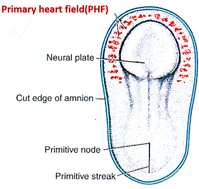
Q. Where is the secondary heart field located? Which parts of the heart develop from primary and secondary heart fields?
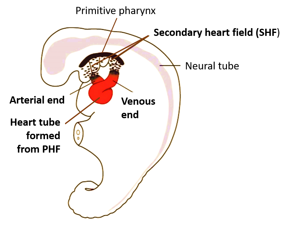
A. The cells forming secondary heart field (SHF) are located in splanchnic mesoderm of the pharynx .
- Cells of PHF will form the atria, left ventricle, and part of the right ventricle.
- Cells of SHF contribute to formation of the outflow tract (arterial end) and sinus venosus (venous end).
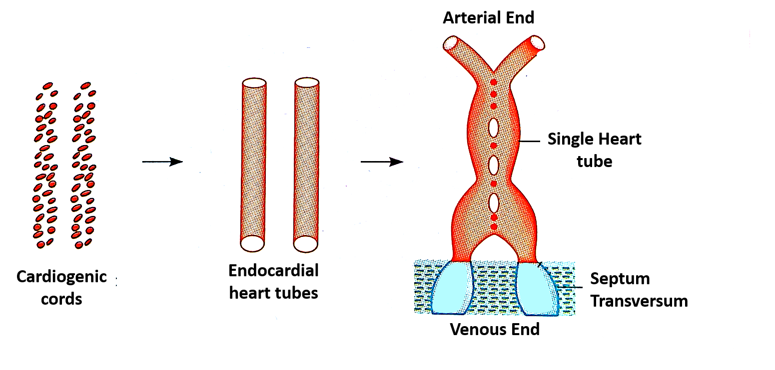
Q. How and where heart tube is formed?
A. Initially two endocardial heart tubes are formed in the cardiogenic area , which later fuse in craniocaudal sequence to form a sing endocardial heart tube.
Q. What is myoepicardial mantle and what are its adult derivatives?
A. The mesenchyme surrounding the endocardial heart tube forms myoepicardial mantle, which subsequently forms myocardium and epicardium (visceral layer of serous pericardium).
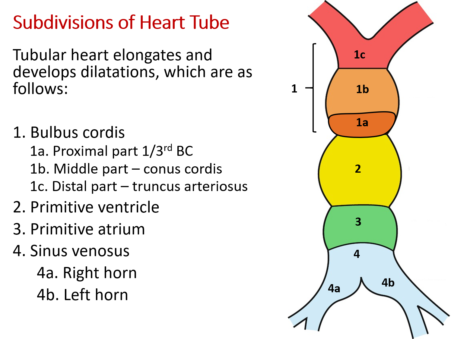
Q. what are the subdivisions of heart tube?
Q. When does the cardiac loop formed?
A. Rapid growth of heart tube results in formation of ‘ U’ and then ‘S’ shaped Cardiac loop during 23-28 weeks.
Q. what are the adult derivatives of various subdivisions of heart tube?
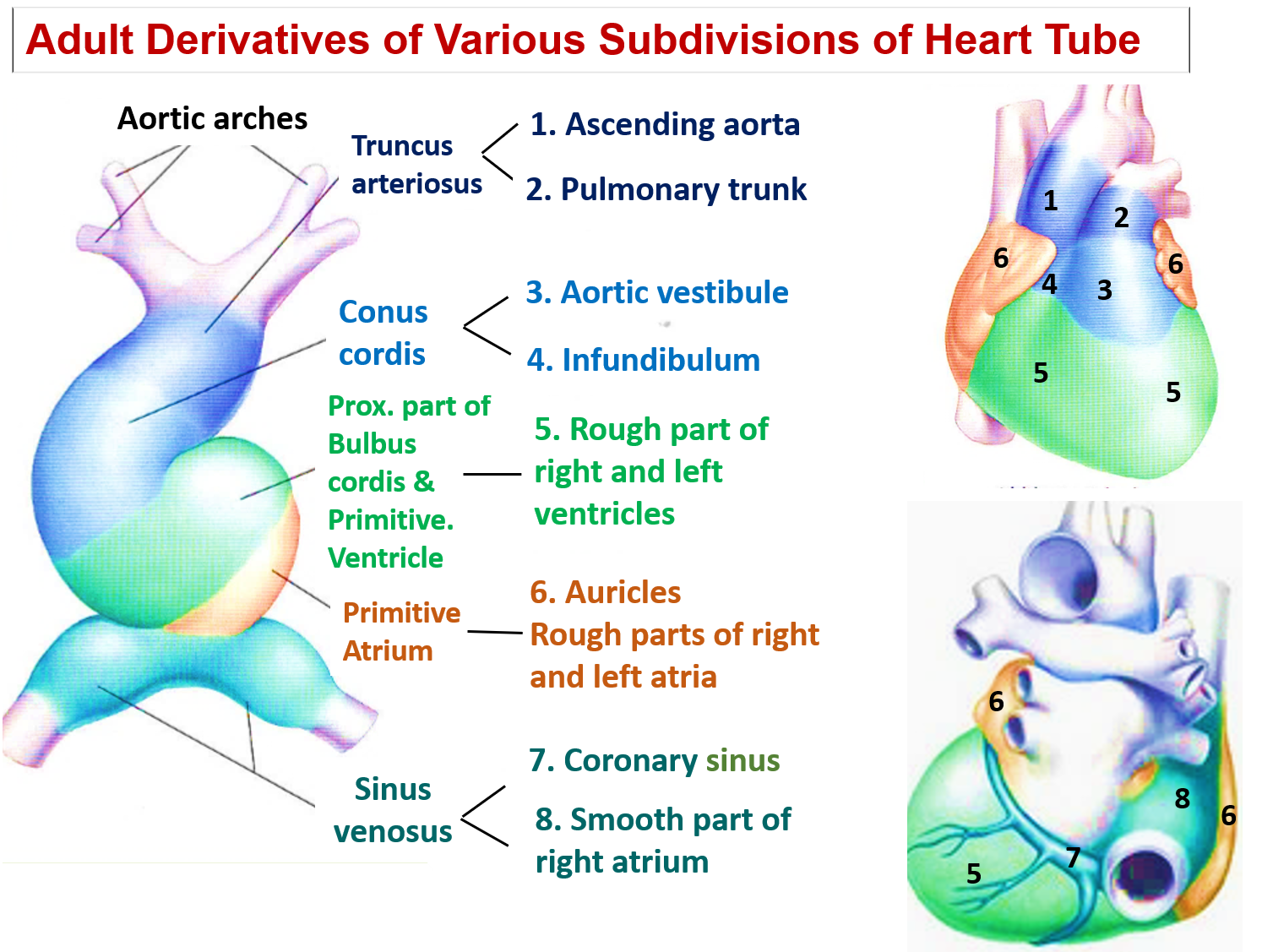
Q. What is the fate of sinus venous?
A. Right horn of sinus venosus enlarges and forms the smooth part of the right atrium. Left horn forms coronary sinus.
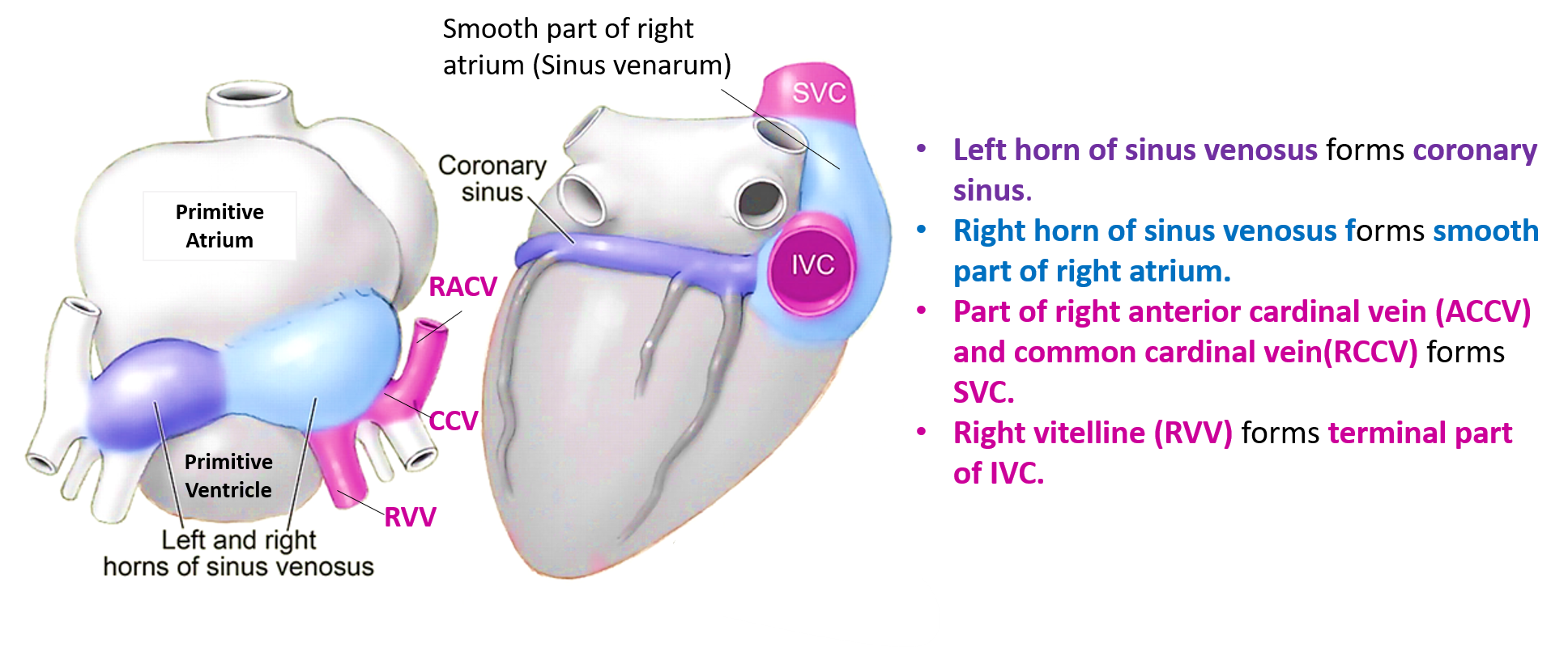
Q. Name the veins that open into each horn of sinus venosus.
A. Each horn of sinus venosus receives:
- Umbilical vein from chorion.
- Vitelline vein from the yolk sac.
- Common cardinal vein from the embryo.
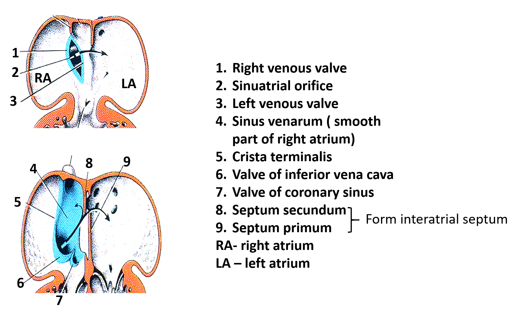
Q. What are the adult derivatives of the right and left valves of sinu-atrial orifice?
A. The right venous valve is divided into three parts and forms:
- Crista terminalis
- Valve of inferior vena cava
- Valve of coronary sinus
Left valve fuses with developing interatrial septum.
Q. Identify the structures labelled 1-9.
Q. Name the embryological components that contribute to the formation of right atrium.
A. Following are the embryological components that contribute to the formation of right atrium.
- Right horn of sinus venosus forms the smooth part of the right atrium, the sinus venarum .
- The muscular part and the the auricle, is derived from the primitive atrium.
- The 2 parts are separated internally by the crista terminalis and externally by the sulcus terminalis.
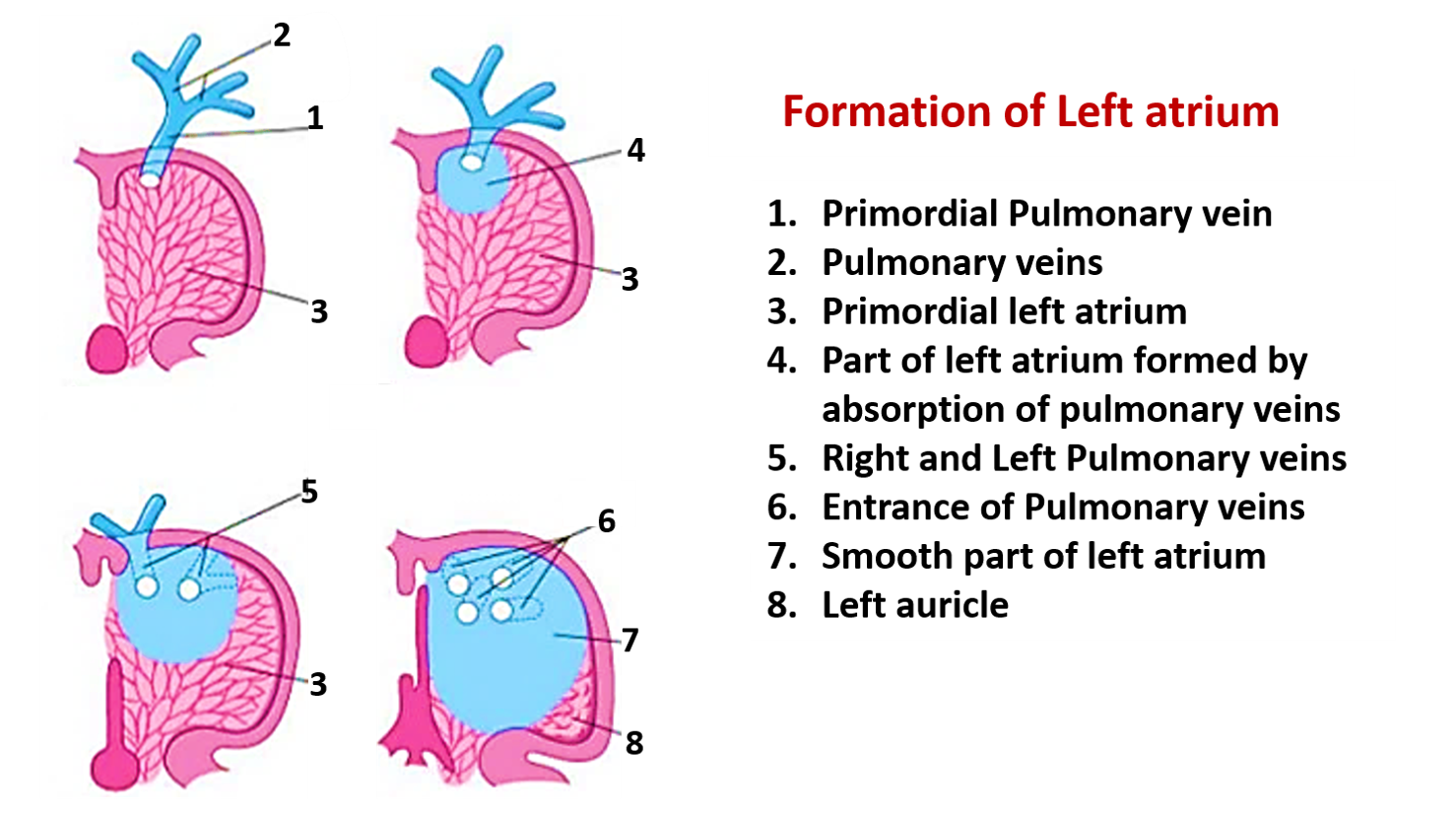
Q. Name the embryological components that contribute to the formation of left atrium.
A. Following are the embryological components that contribute to the formation of right atrium.
- The primitive pulmonary vein and its 4 main branches become partially incorporated into the left atrium and form its smooth part
- The part derived from the primitive atrium retains a trabeculated appearance & forms left auricle
Q. Identify the structures labeled 1-8.
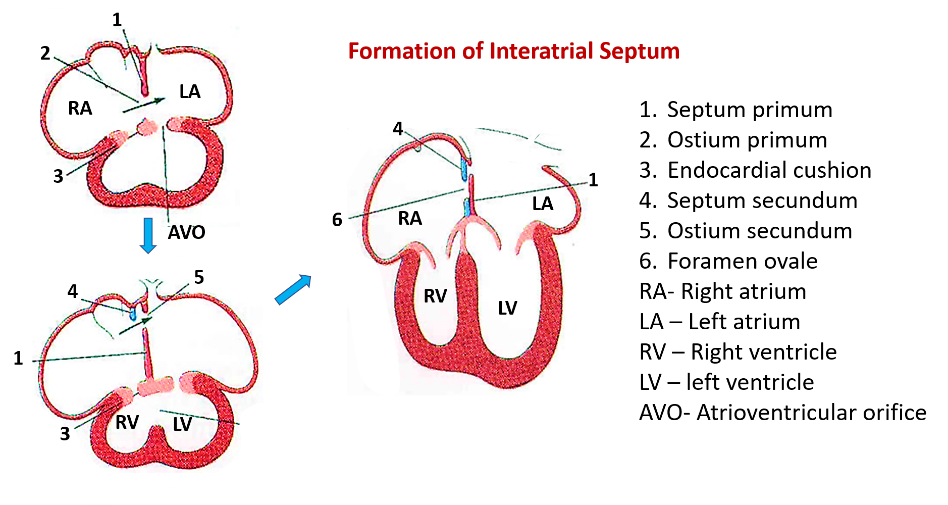
Q. Describe formation of interatrial septum.
A. Formation of interatrial septa :
- Involves formation of two two septa i.e.Septum primum and septum secundum.
- Both are produced by proliferation of the endothelium.
- Septum primum develops from dorsocranial atrial wall of primitive atrium & grows toward endocardial cushions. Space between septum primum & endocardial cushions is called ostium primum.
- Septum primum contacts endocardial cushions & foramen primum is obliterated.
- Septum secundum grows down from roof of atrium to the right of septum primum.
- Septum Secundum covers foramen secundum, but an oblique foramen ovale remains patent.
- In the fetus blood can flow from the right atrium through the foramen ovale.
- At birth the two septa become pressed against each other due to increased pressure in the left atrium. They eventually fuse.
Q. Identify the structures labeled 1 – 6.
Q. What are the adult derivatives of septum primum and septum secundum?
A. In Adults:
- The inferior edge of the septum secundum is crescent shaped and called the limbus fossa ovalis.
- Septm primum forms fossa ovalis.
Q. What is the embryological basis of patent foramen ovale?
A. Patent foramen ovale is a flaplike opening between the right and left atria after birth at the location of the fossa ovalis . It occurs due to non-fusion of Septum primum and secundum .
Q. Describe the formation of interventricular septum.
A. Interventricular septum formation starts by end of 4th week.
- As the ventricles grow and expand laterally, medial walls fuse to forms the muscular Interventricular septum .
- Communication between the ventricles below endocardial cushions and above the muscular interventriclular septum is closed by membranous interventriclular septum at end of 7th week.
Q. Name the embryological components that contribute to the formation of membranous interventricular septum.
A. Following are the embryological components that contribute to the formation of membranous interventricular septum.
- Right and left bulbar ridges (from neural crest)
- Endocardial cushions.
Q. Identify the structures labeled 1 – 8.

Q. What is dextrocardia?
A. Location of heart in the right side of chest.
- Atria and ventricles are transposed.
- Heart tube bends in reverse direction i.e. to the left.
- Could be a part of situs inversus in which there is complete reversal of all organs.
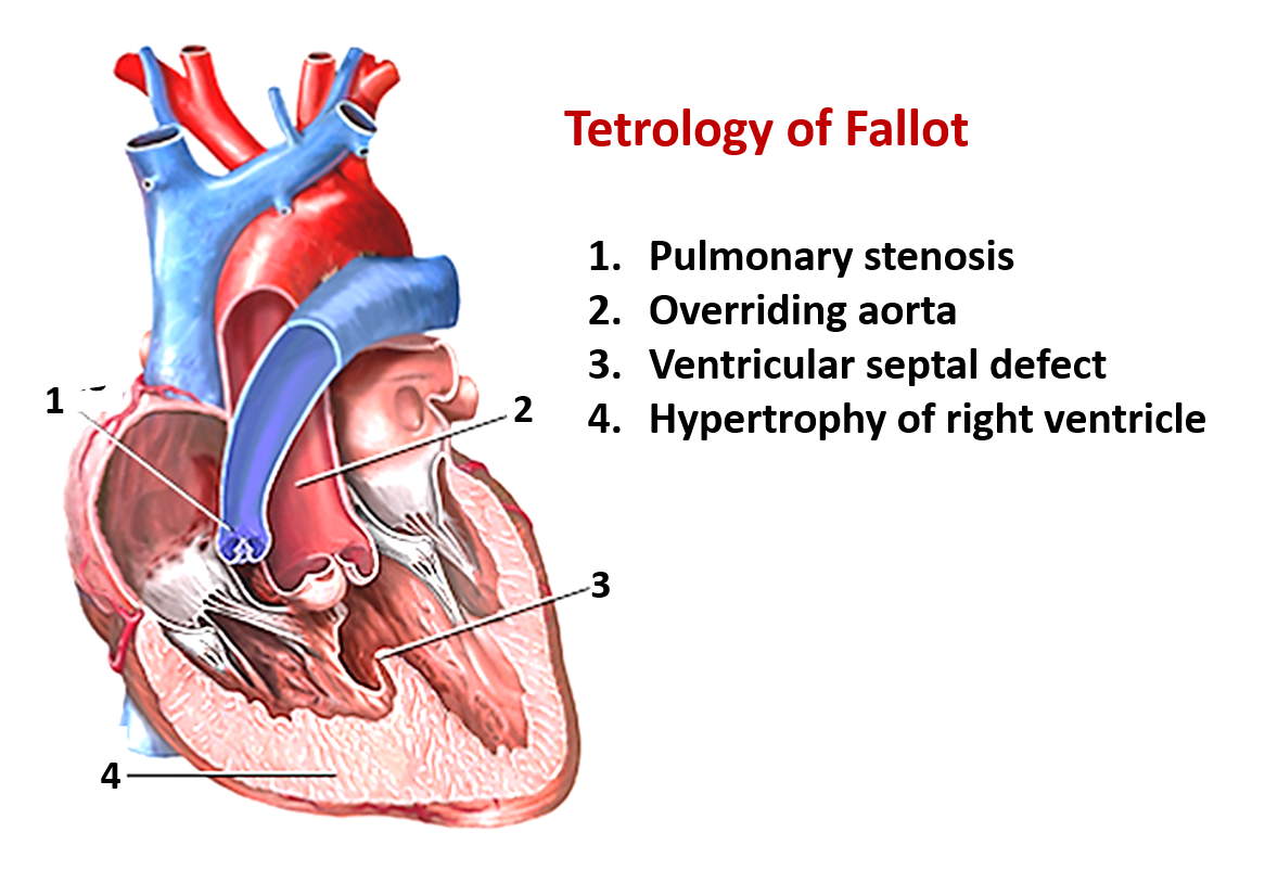
Q. What are the abnormalities in ‘Tetrology of Fallot’?
A. Four abnormalities occur due to anterior displacement of conotruncal septum which results in unequal division of conus. the four defects are:
- Pulmonary stenosis
- Interventricular septal defect
- Overriding aorta
- Hypertrophy of right ventricle

Please we need development of all systems
I am so soo grateful about this website. Absolutely beautiful content. I prepared my total anatomy from this for university exams. Covered the whole syllabus perfectly. Very much grateful to you guys. Thank you so muchhhh. Can’t thank you enoughhhh. I wish we had the same site for other subjects too because am totally dependent on this. So try about other subjects too
how did you do?
This was so helpful. Thank you
Thanks a lot, kindly fix the quizes on thorax anatomy
This is so helpful. Thank you so much.
Please we need development of the whole Gastrointestinal system fully detailed like this one. Thank you.
God bless you abundantly