Bone is a specialised connective tissue, its matrix is impregnated with calcium salts (mainly calcium hydroxy-apatite [CA10(PO4)6(OH)2]. It is highly vascular and has great regenerative power. There are total of 206 bones in adults.
Functions of Bones
Following are the functions of bone:
• They form skeletal framework of the body and give shape to the body
• They protect vital organs such as brain, heart and lungs.
• They store mineral salts e.g. calcium and phosphorus.
• They are the sites of blood cell production.
• They provide surface for attachment of muscles, thereby help in producing movements.
• Some of the bones of skull have air sinuses which add resonance to the voice.
Classification of Bones
Bones are classified based on:
- Shape
- Structure
- Ossification
- Region
Classification of Bones- according to shape
1. Long bones
- Typical long bones:
- Their length exceeds the breadth.
- They are weight bearing.
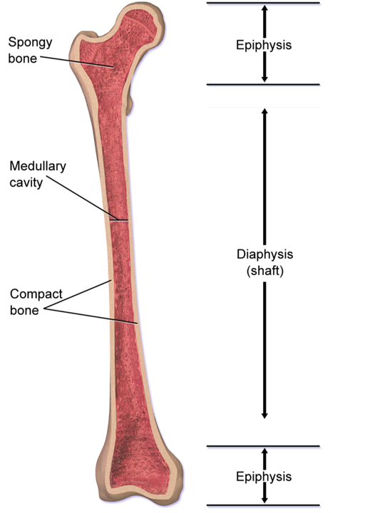
- A long bone presents a shaft and two ends.
- The shaft ossifies from primary center of ossification (appears before birth) and ends ossify from secondary centers of ossification (usually appear after birth, except lower end of femur and upper end of tibia, where secondary center of ossification appears just before birth).
- The shaft consists of tube of compact bone which contains medullary cavity filled with bone marrow.
- The shaft usually has 3 surfaces separated by 3 borders.
- They ossify in cartilages.
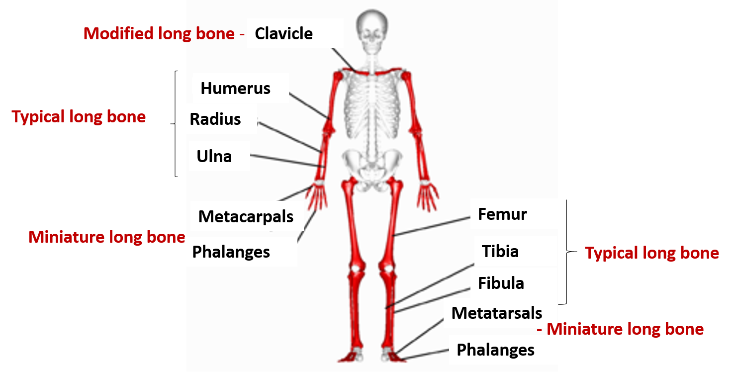
- Examples – humerus, femur, radius, ulna, tibia, fibula.
- Miniature long bones: have a shaft and one epiphysis, e.g. Metacarpals, metatarsals and phalanges. They are mostly confined to limbs.
- Modified long bones: have no medullary cavities, e.g. clavicle and body of vertebra.
2. Short bones
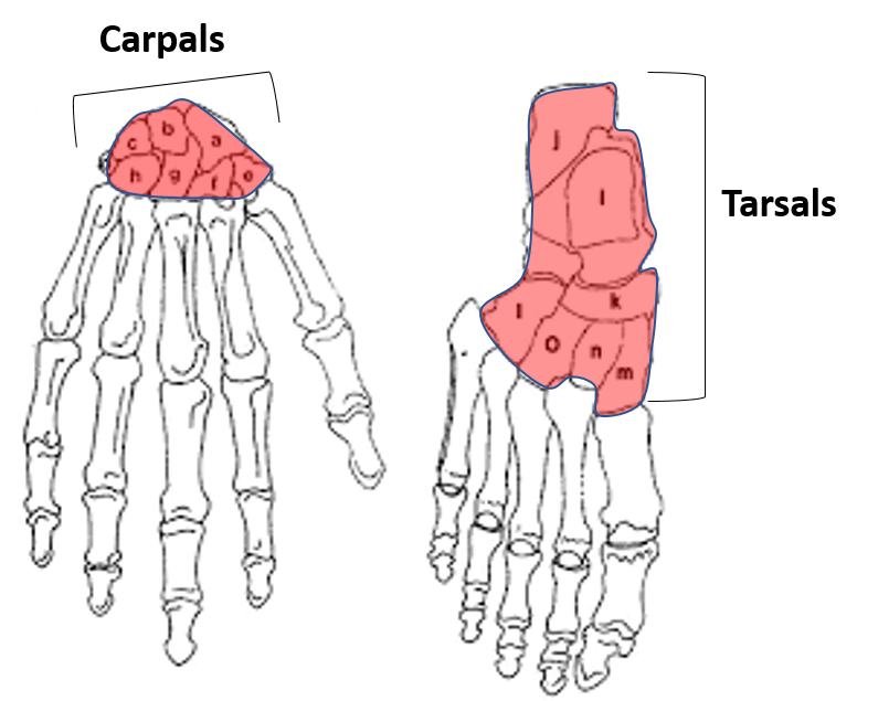
- Their shape is usually cuboid.
- They have a central marrow cavity which is surrounded by spongy bone and a plate of compact bone outside
- They ossify in cartilage.
- They ossify after birth (except talus, calcaneus and cuboid where ossification begins before birth).
- Examples – carpals and tarsals.
3. Flat bones
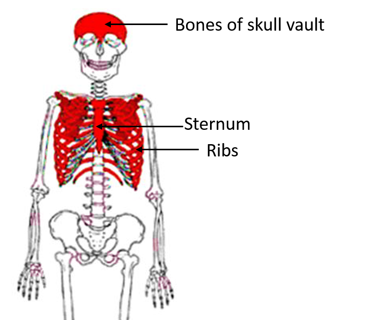
- Consist of two plates of compact bone with intervening spongy tissue (in skull bones it is called diploe).
- Examples: Bones of vault of skull (frontal, parietal), ribs sternum.
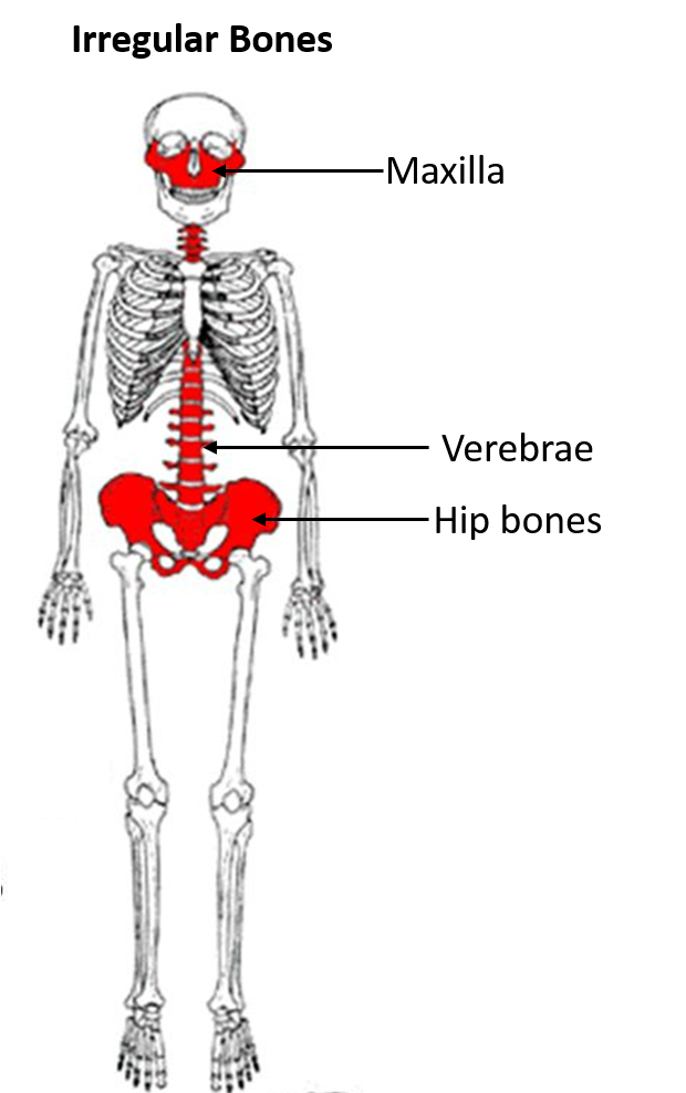
4. Irregular bones
- They are irregular in shape.
- Examples Hip bone, vertebra, bones of base of skull (sphenoid, maxilla, ethmoid).
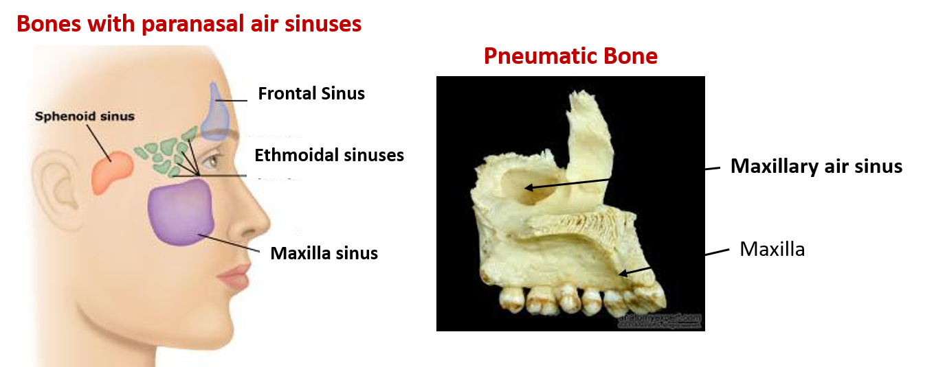
5. Pneumatic bones
- These bones contain air filled spaces (paranasal air sinuses) that are lined by mucous membrane.
- They are present around the nasal cavity.
- They help in adding resonance to the voice.
- Examples: Maxilla, sphenoid, ethmoid, frontal.
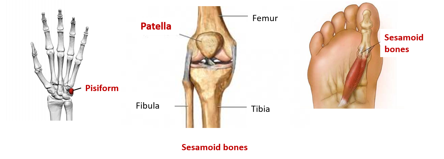
6. Sesamoid bones
- Examples: patella, pisiform, fabella.
Characteristic features and functions of sesamoid bone:
Sesamoid bones are the bones that are embedded in the tendons e.g. patella in the tendon of quadriceps femoris and pisiform in the tendon of flexor carpi ulnaris. They have the following characteristic features:
- They develop in the tendons and ossify after birth.
- They are found where the tendons cross the end of long bones of limbs.
- They are not covered by periosteum.
- They don’t have Haversian system.
- Their surface that comes in contact with the bone is covered by hyaline cartilage.
Functions of sesamoid bone:
• To minimize friction.
• To alter the direction of pull of a muscle.
• To maintain local circulation.
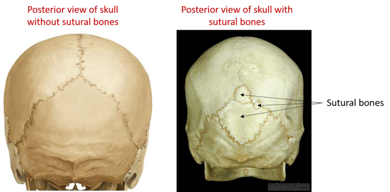
7. Accessory or supernumerary bones
These bone are not regularly present.
Are commonest in skulls (sutural or Wormian bones).
They may be mistaken for fractures.
Classification of Bones- according to the structure
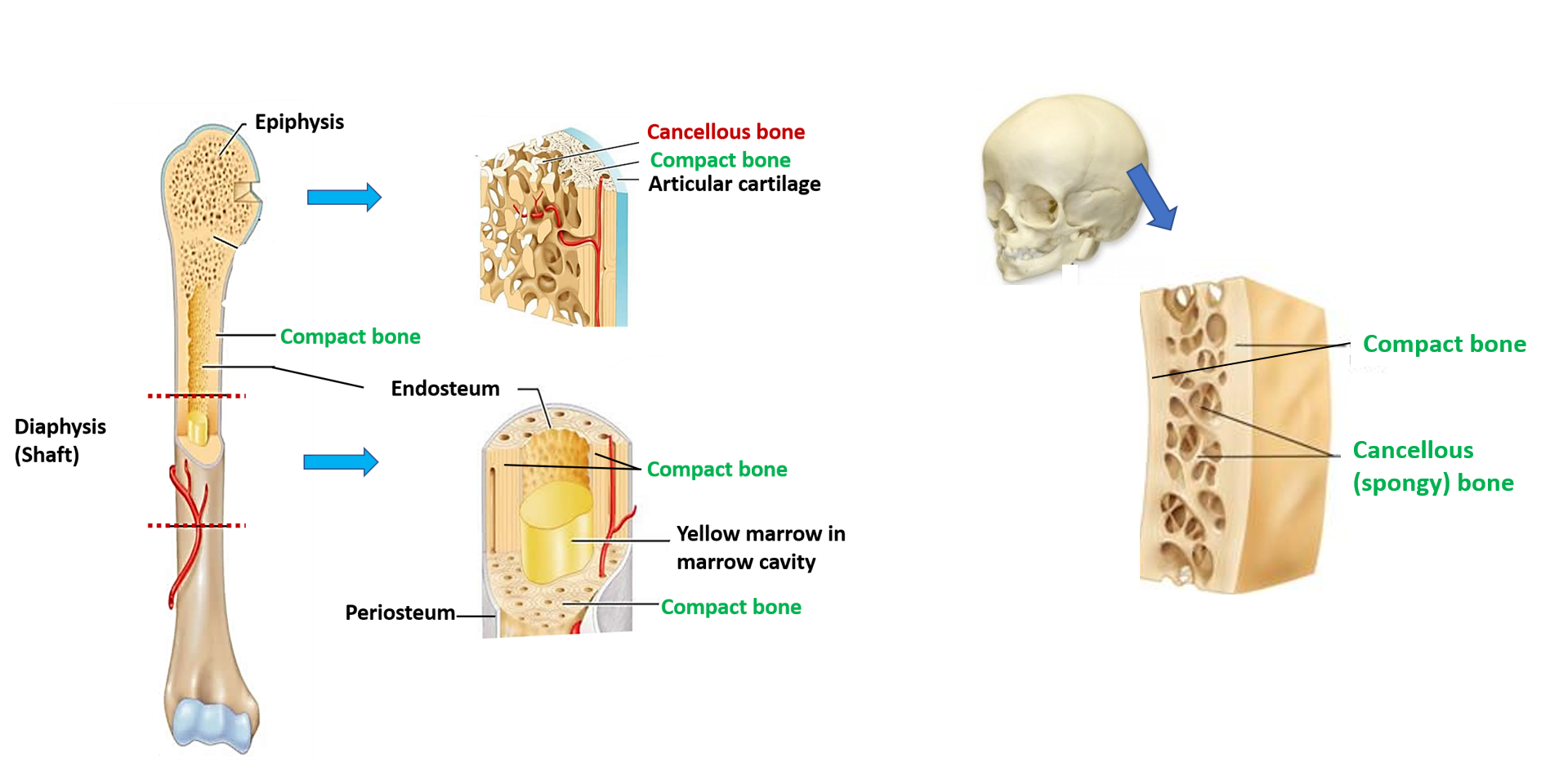
- Compact bone: are dense in texture like ivory: e.g. shaft of the long bones.
- Cancellous bone (spongy or trabecular): are made up of bony trabeculae between which marrow space are present. e.g. ends of long bones.
Classification of Bones- according to the ossification
- Membranous bones: bones are formed by intramembranous ossification, they ossify in mesenchymal condensation. e.g. bones of vault of skull ( parietal, frontal & occipital) and bones of face.
- Cartilaginous bones: bone are formed by endochondral ossification, i.e they ossify in cartilage. e.g. Bones of limbs (humerus, femur, radius, ulna, fibula,tibia), bones of base of skull, vertebrae, ribs, sternum.
- Membrano-cartilaginous bones: Some bones partly develop in membrane and partly in cartilage e.g. mandible, clavicle, occipital, temporal and sphenoid.
Classification of Bones- Regional
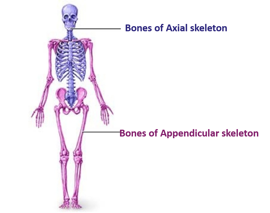
• Axial bones: bones of skull, vertebrae, ribs and sternum.
• Appendicular bones: bones of upper (clavicle, scapula, humerus, radius and ulna, carpals, metacarpals and phalanges) and lower limb (hip bone, femur, tibia, tarsals, metatarsals and phalanges).
Parts of Developing Long Bone
Following are the parts of a developing long bone are: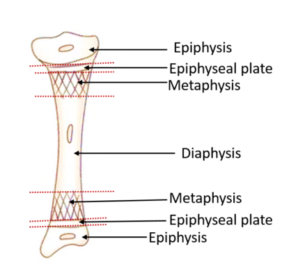
Epiphysis: The ends of long bones that ossify from the secondary centre of ossification are called epiphysis. Epiphyses are made of spongy bone covered by a thin layer of compact bone.
Diaphysis: The elongated, cylindrical shaft of long bone that ossifies from the primary centre of ossification. It is made up of compact bone and encloses a tubular cavity called marrow cavity.
Metaphysis: It is the part of diaphysis that is adjacent to the epiphyseal plate. This is the most active site of bone formation in the developing bone. It has rich blood supply.
Epiphyseal plate: it is a thin plate of hyaline cartilage placed between the diaphysis and epiphysis. The continuous proliferation of cartilage cells in the epiphyseal plate is responsible for the growth of developing bone in length. The epiphyseal plate disappears when the growth in the length of bone stops.
Parts of an Adult Long Bone
Parts of an adult long bone: Adult long bone has two ends and an intervening shaft.
• Shaft (diaphysis)The shaft of the long bone is composed of the following from outside to inside
• Periosteum: It is a thick fibro-cellular layer that covers the outer surface of bone except the articular surfaces which are covered by articular cartilage (hyaline cartilage). It has two layers:
- Outer fibrous layer
- Inner cellular layer or osteogenic layer (containing osteoprogenitor cells which form osteoblasts).
- It is united to the underlying bone by Sharpey’s fibers (collagen fibers).
- It has very rich nerve supply and therefore it is the most pain sensitive part of bone.
• Cortex (cortical bone): It is made up of dense compact/cortical bone.
• Marrow cavity: deep to the cortex is the medullary cavity. It is lined by endosteum and is filled with bone marrow (depending upon age of the individual it can be red or yellow marrow).
• Ends of long bone (Epiphysis): Are made up of cancellous bone (having bony trabeculae and marrow spaces (filled with red bone marrow)). The articular surfaces at the ends are covered by articular (hyaline) cartilage).
b. Bone marrow: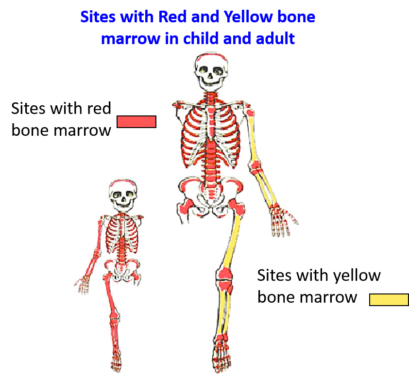
• Is a loose vascular tissue.
• It is present in the medullary cavity of the long bones and marrow spaces of spongy bones.
• It is the site of haemopoiesis.
• There are two types of bone marrow:
o Red bone marrow
o Yellow bone marrow
• At birth the red bone marrow is present in all the bones, it is an active site of hemopoiesis (blood cell formation).
• As the age advances the red marrow is replace by yellow bone marrow in the medullary cavity of long long bones.
• In adults, the red marrow is found only in the spongy bones.
• The sites where red bone marrow is found in adults are:
o Iliac crest of hip bone
o Sternum
o Ribs
o Vertebrae
o Ends of long bones
o Skull bones
Types of Epiphysis
Q. Enumerate the types of epiphyses and give one example of each.
A. Types of epiphyses:
1. Pressure epiphysis:
o Is present at the end of the long bone.
o Is articular in nature.
o Takes part in transmission of weight.
o Examples: head of humerus and femur, lower end of radius.
2. Traction epiphysis:
o Is produced due to the pull of the muscle and therefore provide attachment to the muscle/s.
o Is non-articular
o Is not involved in transmission of weight
o Examples: greater and lesser tubercles of humerus, greater and lesser trochanters of femur and mastoid process of temporal bone.
3. Atavistic epiphysis:
• Is phylogenetically an independent bone in lower animals but in humans is part of another bone.
• Examples: coracoids process of scapula, posterior tubercle of talus.
4. Aberrant epiphysis:
• Is not always present but may be present in some individuals.
• Examples: epiphysis at the head of first metacarpal and base of other metacarpals.
* Normally metacarpals have only one epiphysis i.e. base of 1st metacarpal and heads of other metacarpals.
Pressure epiphyses ossify earlier than the traction epiphyses i.e. secondary center of ossification appear earlier in pressure epiphyses than in traction epiphyses.
Arterial Supply of Developing Long Bone
Q. Describe the arterial supply of developing long bone.
A. Developing long bone is supplied by the following arteries:
• Nutrient artery:
o It enters the shaft through the nutrient foramen.
o Runs obliquely through the cortex of shaft.
o Reaches medullary cavity and divides into ascending and descending branches.
o These branches reach the metaphysis and form ‘hair pin loops’ and anastomose with the metaphyseal and epiphyseal arteries.
o The nutrient artery supplies the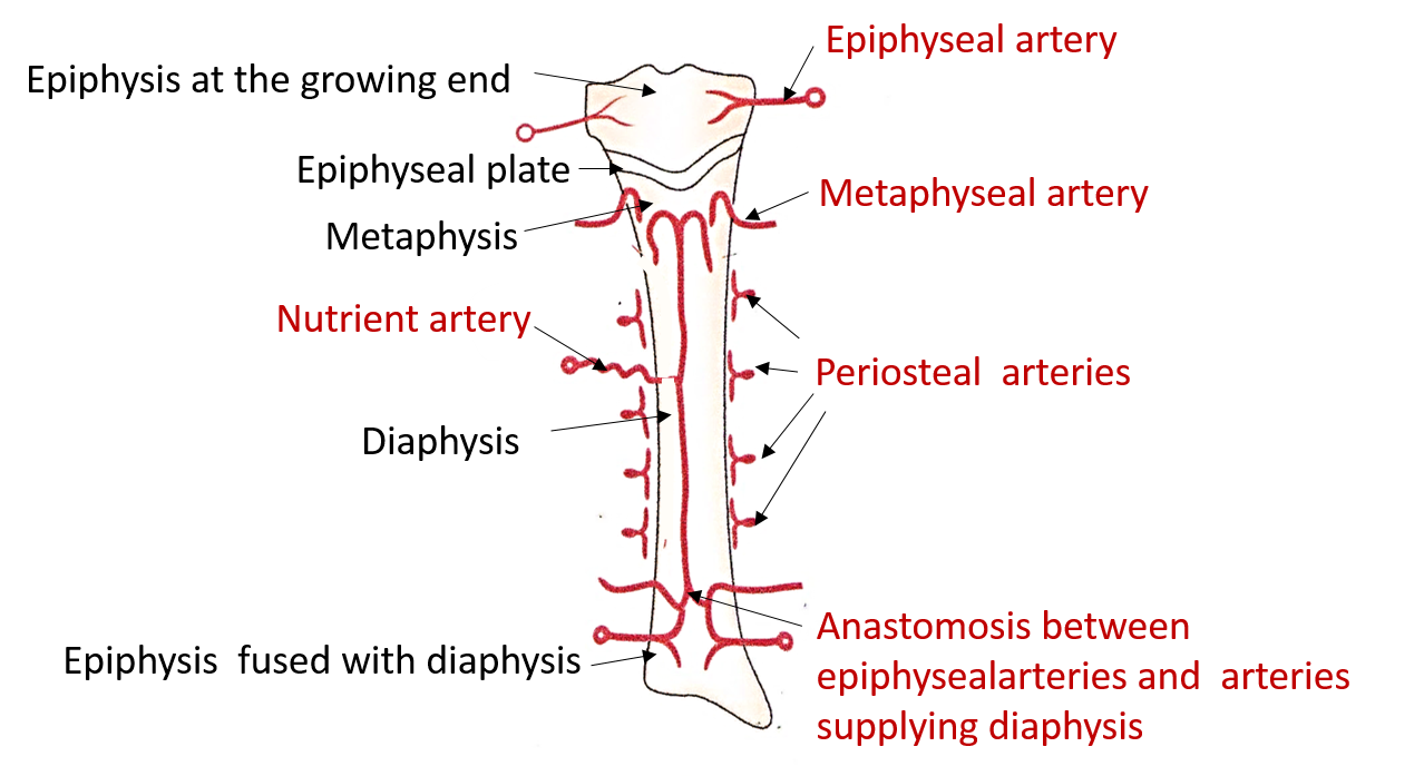
medullary cavity
inner 2/3rd of cortical bone of diaphysis
metaphysis
• Periosteal arteries:
o Are branches of neighbouring muscular arteries and therefore they are especially numerous beneath the muscular attachments.
o They ramify beneath the periosteum and reach the underlying cortical bone through the Volkman’s canals.
o They supply periosteum, and outer 1/3rd of the cortical bone of shaft.
• Epiphyseal arteries:
o They are derived from the periarticular (around the joint) anastomosis present around the non-articular surface of ends of long bones.
o They enter the epiphyseal ends through the numerous foramina present in the non articular part. The number and size of these foramina gives an idea of the vascularity of the ends of long bones.
• Metaphyseal arteries:
- Numerous arteries arise from the anastomosis around the joint and pierce the metaphysis along the attachment of the joint capsule
Ossifiction
Q. Describe briefly:
- Ossification centers
- Growing end of the long bone.
- Types of ossification
A. Ossification centers
The process of bone formation is known as ossification. Sites where begins are referred to as centers of ossification. There are two types of centers of ossification:
 Primary center of ossification: is the site in the mid region of shaft of long bone where ossification begins earliest. It forms the main part of the bone (diaphysis in long bones) It usually appears before birth (usually before the end of the fourth month of . intrauterine life).
• Secondary center of ossification: The secondary center of ossification usually appear after birth from which the ends of the developing long bone ossify. They form the epiphyses of the long bones. In case of femur the secondary center of ossification for the lower end appears just before birth
Growing end of the long bone
It is that end of the long bone where the epiphysis fuses with the diaphysis later than the other end (it continues to grow in length a little longer). The growing end of the long bone is opposite to the direction of the nutrient foramen (exception to the rule is fibula where the nutrient foramen is directed towards the growing end).

Types of Ossification
Bone can be formed in two ways:
• Intramembranous ossification
• Intracartilagenous or endrochondral ossification
Membranous ossification: In this type of ossification, the mesodermal model is directly converted into bone by mineralization of the matrix. Most of the flat bones such as frontal and parietal bones , parts of occipital, temporal, mandible, maxilla and clavicle bones are formed by intramembranous ossification It can be divided into the following 4 stages:
1. Mesenchymal condensation and appearance of centers of ossification: one or two centres of ossification appear in the condensed mesenchyme. The center of ossification is characterised by excessive vascularity and differentiation of mesenchymal cells into osteoblasts.
2. Formation of osteoid matrix and its calcification: osteoblasts which secrete gelatinous uncalified matrix and collagen fibers (osteoid). Calcium salts are deposited in the osteoid. The osteoblasts trapped in mineralized matrix become osteocytes.
3. Formation of spongy ( trabecular) bone: bony lamellae are formed around the blood vessels and are arranged in the form of trabeculae which are separated by spaces containing blood cells ( marrow spaces). Thus spongy bone is formed.
4. Formation of compact bone and periosteum: the osteoblasts at the periphery lay down concentrically arranges bony lamellae, forming osteons (Haversian system) of compact bone ( spongy bone is replaced by compact bone). Outside the compact bone mesenchymal cells form fibrous periosteum with osteoblasts on inner surface.

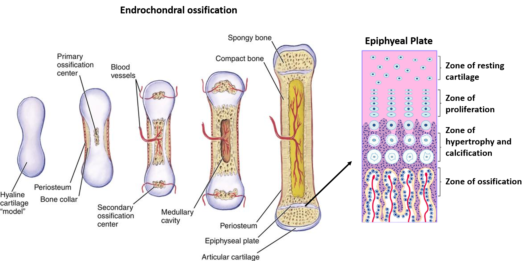
Endrochondral ossification: In this type of ossification, firstly a hyaline cartilage model of bone is formed, which is later replaced by bone. It involves the following 4 stages:
1. Chondrification: mesenchymal cells differentiate into chondroblasts, which form a hyaline cartilage model for the future bone. At he periphery mesenchymal cells for perichondriumwhic contains osteogenic cells on the inner surface.
2. Appearance of centres of ossification: one or more centres of ossification appear in the cartilage model, chondrocytes are first hypertrophied and later they degenerate. matrix is reduced to thin partitons , which later get calcified.
3 .Formation of Periosteal bone collar :once the matrix is calcified, the perichondium is now called periosteum. The osteoblasts on the inner surface of periosteum form a layer of bone beneath the periosteum called periosteal collar of bone.
4. Invasion of periosteal sprouts and osteogenesis: Periosteal sprouts arise from deeper layer of periosteum (contain blood vessels, osteoblasts and osteoclasts) and invade the underlying calcified matrix. The osteoclast resorb the matrix to form large interconnecting spaces called marrow spaces which ultimately join together to form medullary cavity. Formation of bone in subperiosteal area of diaphysis continues to increase thickness of the bone ( appositional growth). The growth of bone in length occurs at epiphyseal plate ( interstitial growth).
The epiphyseal plate has five zones:
• Resting zone
• Zone of proliferation
• Zone of hypertrophy
• Zone of calcification and dying chondrocytes
• Zone of ossification
Laws of Ossification
1. Primary center of ossification appears before birth and is usually single. Exception – Carpals and tarsals ( cuneiforms, navicular) ossifiy after birth and clavicle has 2 primary centers of ossification.
2. Secondary centres of ossification appear after birth and can be can be single or multiple. Exception – Lower end of femur and upper end of tibia ( secondary centers at these ends appear before birth, at the end of the 9th month of intrauterine life).
3 The direction of the nutrient foramen is opposite to the direction of the growing end of the bone.
4. The secondary center of ossification for pressure epiphysis appear before that of traction epiphysis (center of ossification in head of humerus ( pressure epiphysis) appears before that in greater /lesser tubercles (traction epiphyses)).
5. The secondary center of ossification that appears first, fuses last with the shaft of long bone. Exception – Lower end of fibula ( secondary center for the upper end appears later but the secondary center for the lower end which appears first fuses earlier with the shaft of fibula.
6. The secondary centers fuse together to form a single epiphysis (compound epiphysis), which in turn fuses with the diaphysis. Exception – Upper end of femur has 3 secondary centers, one each for the head , greater trochanter and lesser trochanter. Each epiphysis fuses independently ( examples of simple epiphysis) with the shaft in the reverse order of appearance i.e the lesser trochanter first followed by about 13 years, the greater trochanter at about 14 years, and the head around 16 years
Applied Aspect
Q. Write the anatomical basis of:
• Cleidocranial dysostosis
• Dwarfism/achondroplasia
• Metaphysis is the common site of osteomyelitis in children
A. Cleidocranial dysostosis: A defect in membranous ossification causes Cleidocranial dysostosis in which ossification of bones ossifying in membrane that is clavicle and bones of skull vault are affected. It is characterised by aplasia of clavicle, increase in transverse diameter of cranium and retardation of ossification of fontanelle.
Dwarfism/Achondroplasia: A defect in endrochondral ossification affects growth and ossification of bones of limbs, which is responsible for dwarfism/achondroplasia in which the limbs are short but the trunk is normal. It may be inherited as autosomal dominant genetic disorder.
Metaphysis is the common site of osteomyelitis in children: Metaphysic is the the area of very active growth of the bone and has rich blood supply. Before fusion of epiphysis with diaphysis i.e. in children, the metaphyseal arteries are end arteries that form ‘hair-pin bends’. The bacteria or infected emboli can be easily trapped in these hair- pin bends of the metaphyseal arteries causing osteomyelitis.
