Advertisements
Introduction
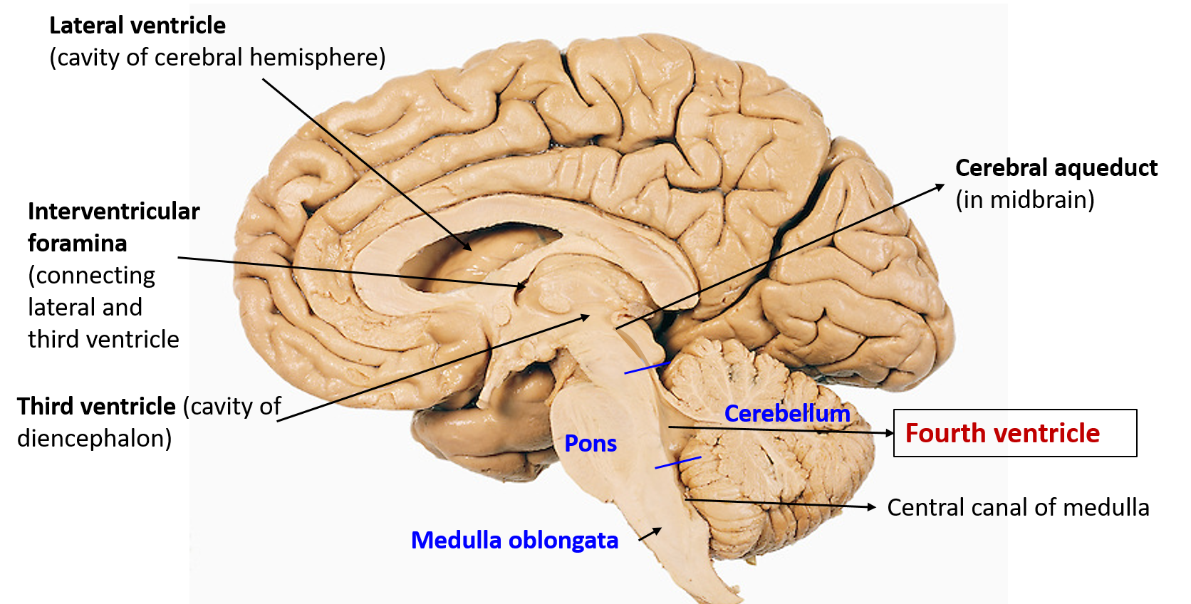
Fourth ventricle is a tent shaped cavity of hind brain lined by ependyma and contains cerebrospinal fluid (CSF) .
Where is fourth ventricle located?
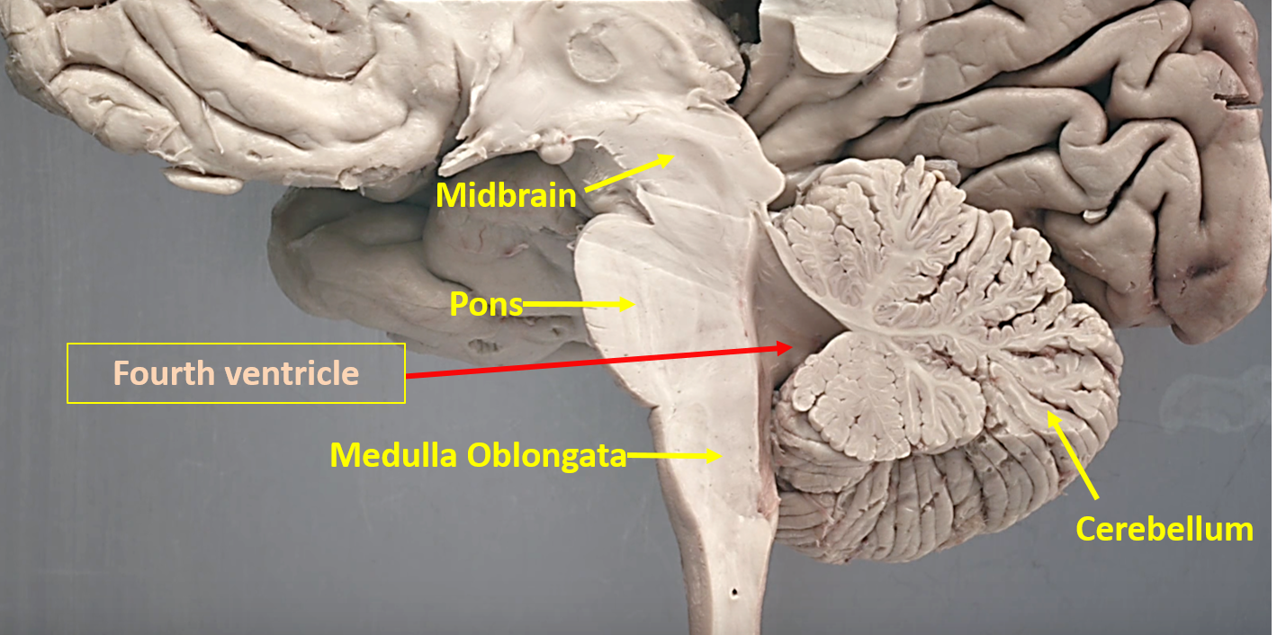
The fourth ventricle is situated dorsal to the pons and upper part of medulla oblongata and ventral to the cerebellum.
What are the boundaries of fourth ventricle?
Fourth ventricle is bounded by
- Two lateral walls
- Roof or dorsal wall
- Floor or ventral wall
Lateral walls of fourth ventricle
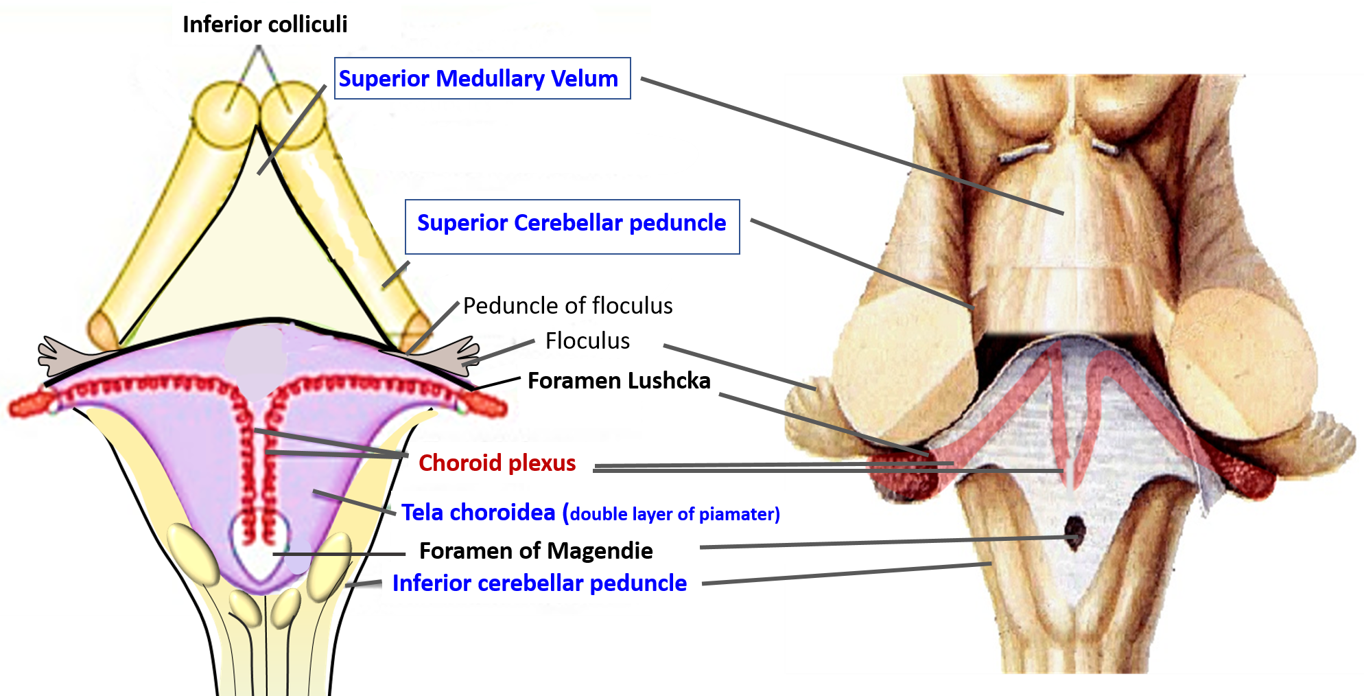
- Upper part of lateral wall is formed by the superior cerebellar peduncle.
- Lower part of lateral wall is formed by the inferior cerebellar peduncle .
Floor or ventral wall of fourth ventricle
- The floor of fourth ventricle is diamond shaped and is also known as rhomboid fossa.
- It is formed by the dorsal surface of pons and upper open part of the medulla.
- At the junction of dorsal part of pons and medulla lie transversely running fibers called stria medullares (fibers in stria medullaris are derived from arcuate nucleus (located anterior to pyramid and are dislodged pontine nuclei) which pass dorsally through the medulla to exit through median sulcus, decussate and pass laterally to enter the inferior cerebellar peduncle to reach cerebellum).
- The floor of fourth ventricle is divided into two symmetrical halves by the median sulcus.

- Each half is further subdivided by sulcus limitans into a medial area called medial eminence and lateral vestibular area.
- In the pontine part of floor of fourth ventricle following features are seen:
- Facial colliculus: medial eminence show an elevation called facial colliculus ( produced by the fibers of facial nerve which form a loop around the dorsal aspect of abducent nucleus).
- Locus ceruleus: at the upper end of sulcus limitans , there is bluish grey area called locus ceruleus ( its neurons produce nor-epinephrine, and may be concerned with paradoxical sleep)
- Superior fovea: below locus ceruleus, sulcus limitance is marked by a depression called superior fovea.
- In the medullary part of the floor of fourth ventricle following features are seen:
- Inferior fovea: A depression at the lower end of sulcus limitance called inferior fovea.
- Hypoglossal and vagal triangles: From the inferior fovea, a sulcus runs obliquely towards the midline and divides median eminence into hypoglossal triangle (medially) and vagal triangle ( laterally). hypoglossal triangle overlies hypoglossal nucleus and deep to vagal triangle is dorsal motor nucleus of vagus.
- Area Postrema: It lies between the vagal triangle and gracile tubercle. Funiculus separans (an ependymal thickening) separates the vagal triangle from the area postrema. it is also known as chemoreceptor trigger zone ( CTZ)/vomitting centre. It is highly vascular and devoid of blood -brain barrier.
Roof of the fourth ventricle
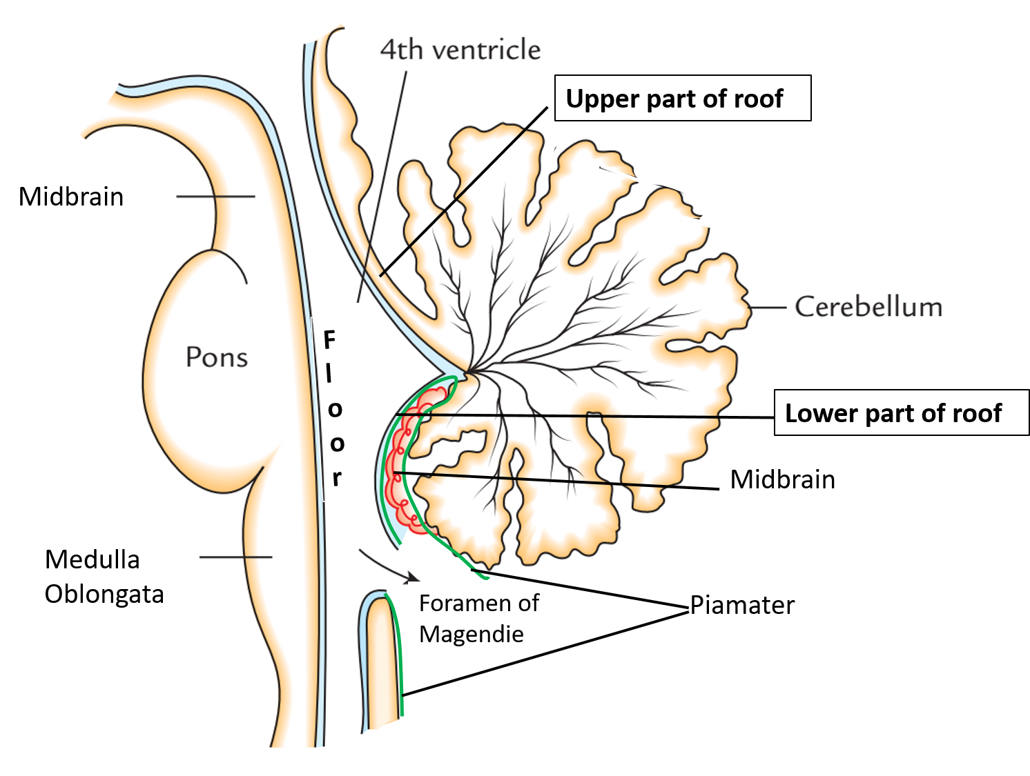
- The roof of fourth ventricle is tent shaped and is divided into upper and lower part.
- In the upper part it is formed by superior medullary velum (white matter between the superior cerebellar peduncles).
- In the lower part i is formed by ependyma covered by double layer of pia matter which forms the tela choroidea of fourth ventricle..In this part there is an aperture called foramen of Magendie which connects the fourth ventricle to cerebellomedullary cistern.
What the structurs/areas with which the cavity of fourth ventricle communicates?
- Superiolyr the fourth ventricle communicates with the cerebral aqueduct.
- Inferiorly with the central canal of lower part of medulla.
- it communicates with subarachnoid space via 3 apertures:
- one median – foramen of Magendie
- two lateral – foramen of Luschka.
Name the recesses of fourth ventricle.
- Two lateral recesses: lie in the interval between the inferior cerebellar peduncle (ventrally) and peduncle of flocculus (dorsally).
- One median dorsal: cranial to nodule (of inferior vermis of cerebellum), extends into the white matter of cerebellum.
- Two lateral dorsal recesses: on either side of the nodule.
Describe the tela choroidea and choroid plexus of fourth ventricle.
Tela choroidea: is a double layer of piamater that forms the lower part of the roof of fourth ventricle.
Choroid plexus:
- Lie between the fold of pia mater forming the tela choroidea.
- There are two plexuses-right and left, each has a vertical limb ( present next to the midline) and a horizontal limb , extending into the lateral recess till the lateral aperture.
- The vertical limbs of the two plexuses are close to each other, so that the whole plexus resembles the shape of letter ‘T’.
Applied Aspect
Internal Hydrocephalus
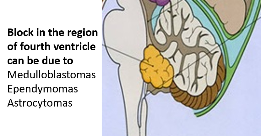
If the opening of fourth ventricle ( foramen of Magendie and Luschka) are blocked (by tumor or adhesions of arachnoid mater), the CSF cannot enter the subarachnoid space from the ventricular cavity. This leads to excessive accumulation in the ventricular system , thereby producing Internal Hydrocephalus.

Very well explained