Introduction
- Cerebrum is the largest part of brain (weighs -`1500 gms ).
- It comprises of two cerebral hemispheres (right and left).
- The two cerebral hemispheres are partially separated by medial longitudinal fissure ( Falx cerebri – the dual folds is located in the median longitudinal fissure. At the bottom of median longitudinal fissure, the two cerebral hemispheres are connected by corpus callosum (commissural fibers connecting same areas of the two cerebral hemispheres).
- The surface of each cerebral hemisphere contains grey matter ( mainly neuronal cell bodies and dendrities), known as cerebral cortex.
- The surface of cerebral hemispheres is irregular as it has elevated folds called gyri which are separated by depressions known as sulci .
- Deep to the cerebral cortex each cerebral hemisphere contains white matter ( mainly myelinated axons).
- Within the white matter are masses of grey matter called basal nuclei/ganglion.
- Each cerebral hemisphere has a cavity filled with cerebrospinal fluid , called lateral ventricle.
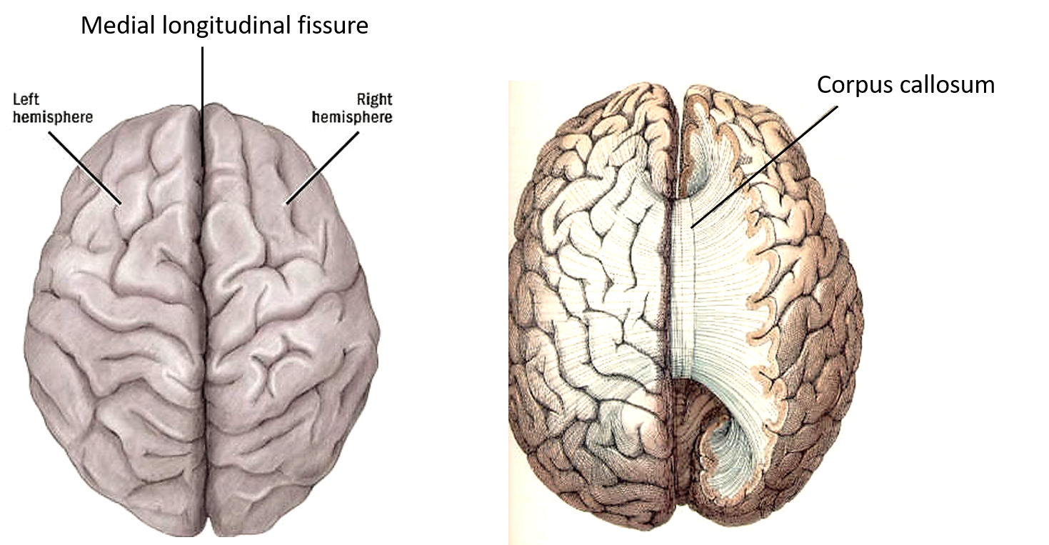
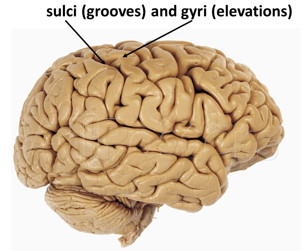
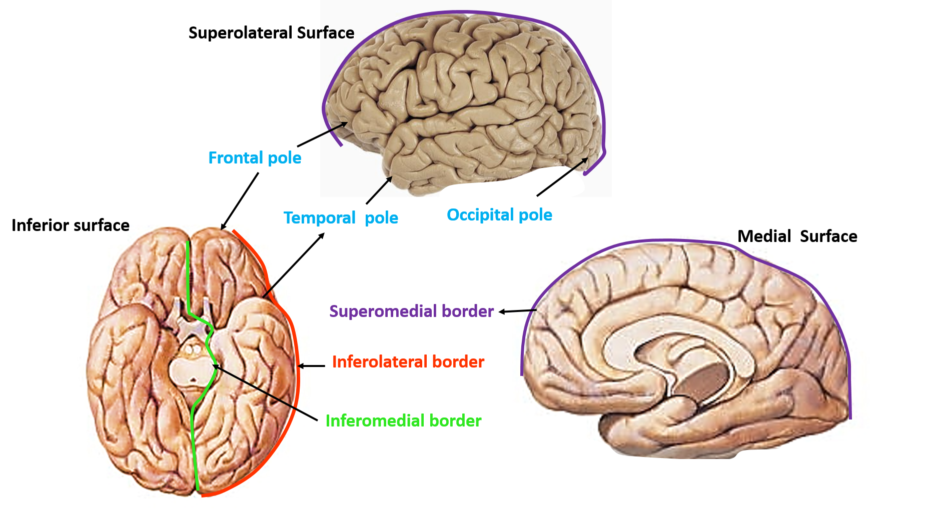
Describe the external features of cerebral hemisphere?
Each cerebral hemisphere has:
- Three surfaces: Superolateral, medial and inferior.
- Three borders: superomedial, inferolateral (orbital part is called supercilliary border), inferomedial ( divided into medial orbital and medial occipital borders).
- Three poles: Frontal, temporal and Occipital.
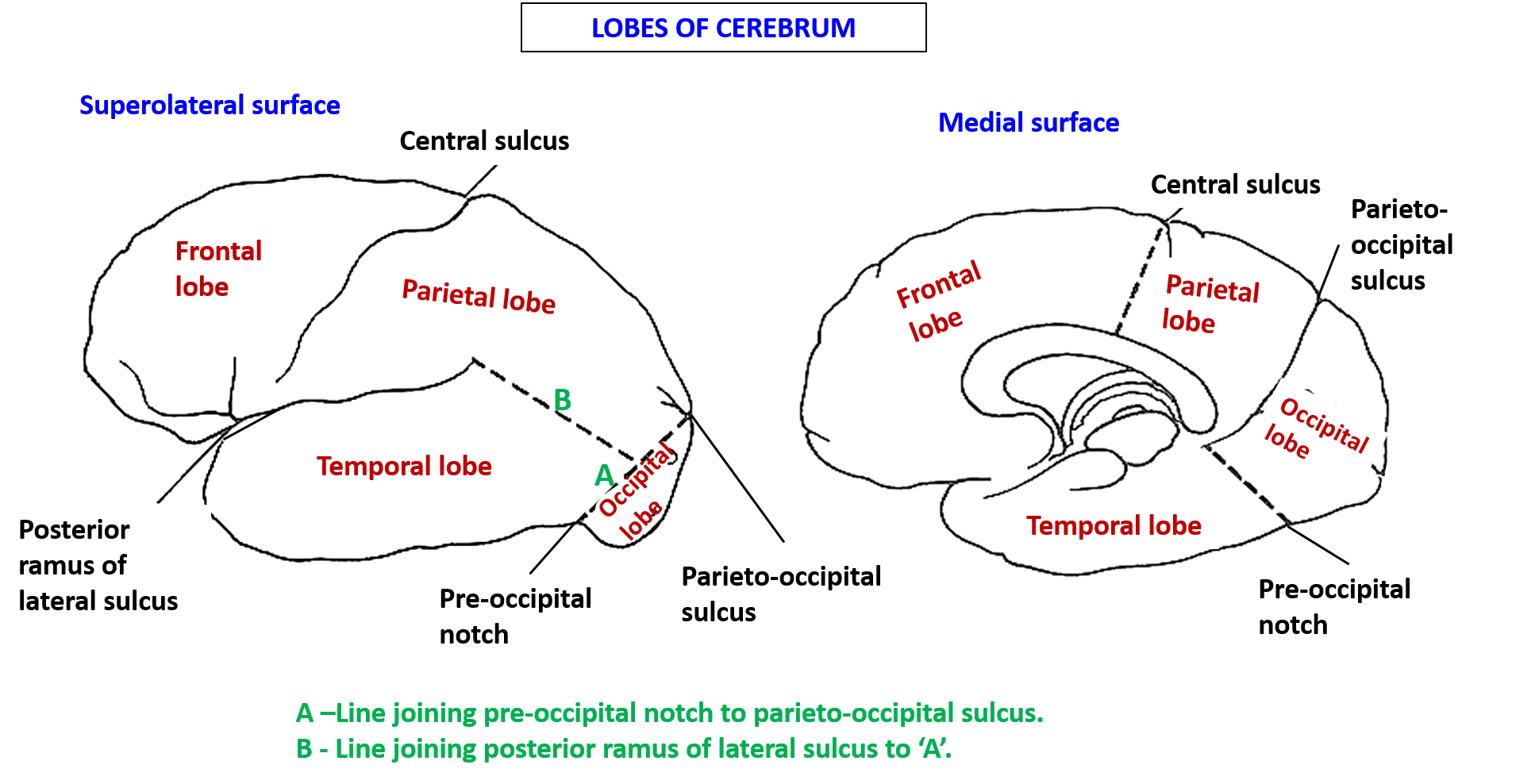
Draw labelled diagram to show the lobes of cerebral hemisphere.
Draw labelled diagram to show the major sulci and gyri of cerebral hemisphere.
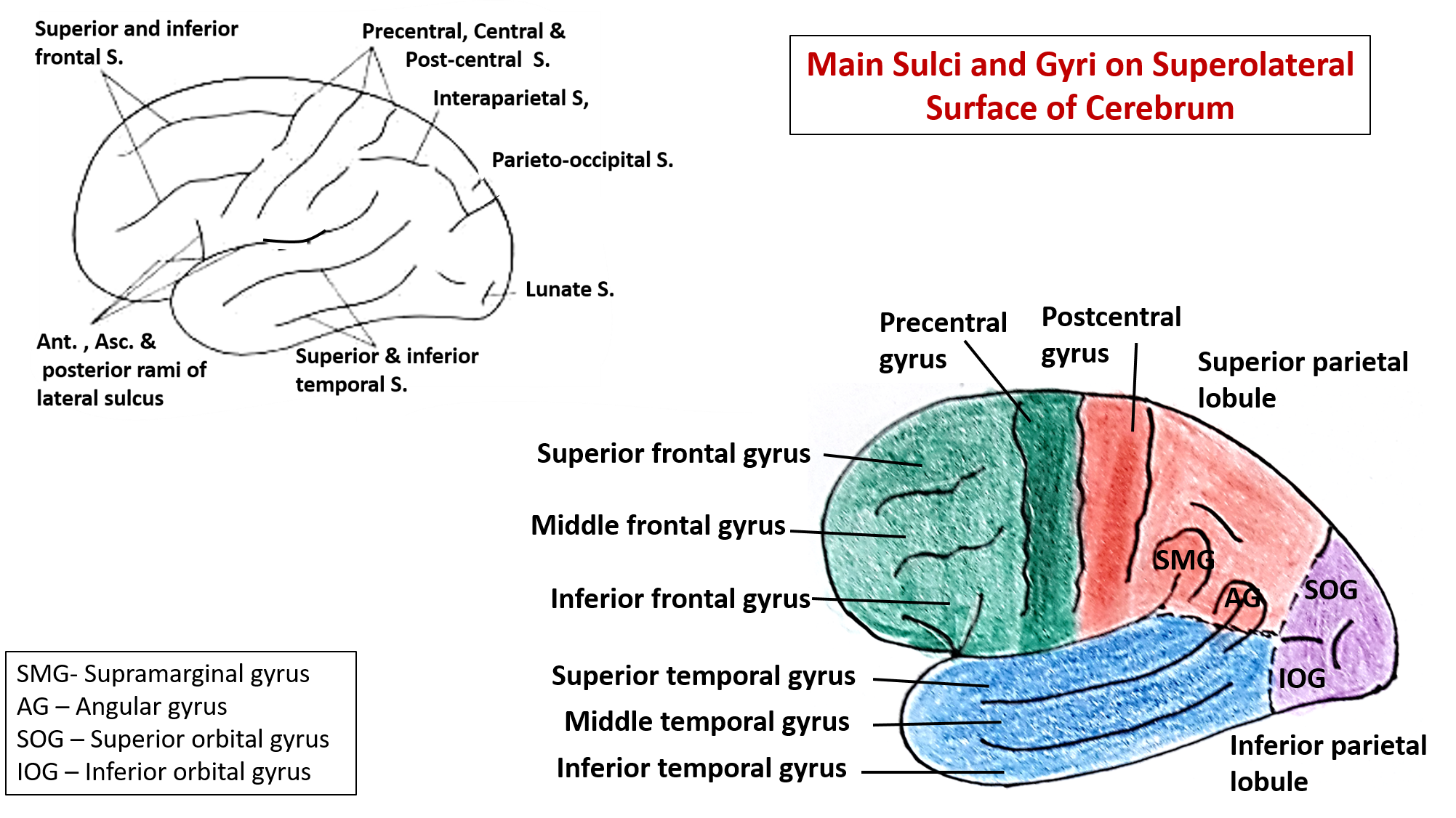
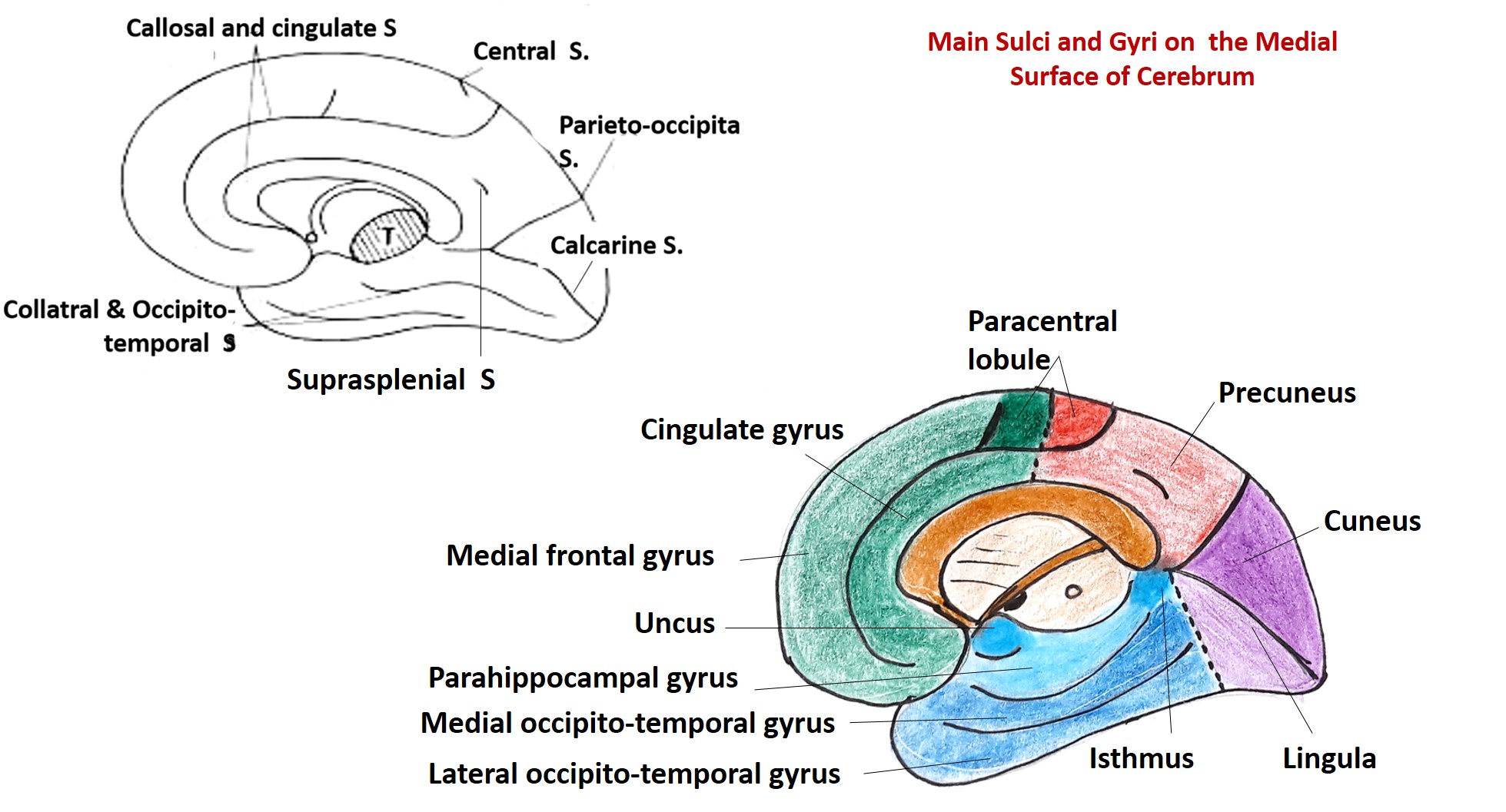
What are functional areas? Draw labelled diagrams to show major functional area of the cerebral hemisphere along with Brodmann numbers.
Various areas of the cerebral cortex perform specific functions. human cerebral cortex has been described to have three types of functional areas:
- Motor areas – control voluntary movement . Mainly present in frontal lobe
- Sensory areas – conscious awareness/perception of general somatic sensations ( pain temperature, touch, proprioception) and special sensations ( vision, hearing, taste, smell).
- Association areas – almost 70% of the cerebral cortex is association cortex. these areas are usually present close to the sensory areas It integrate various type of sensory information and to direct behaviour, communication and intellect.diverse information.
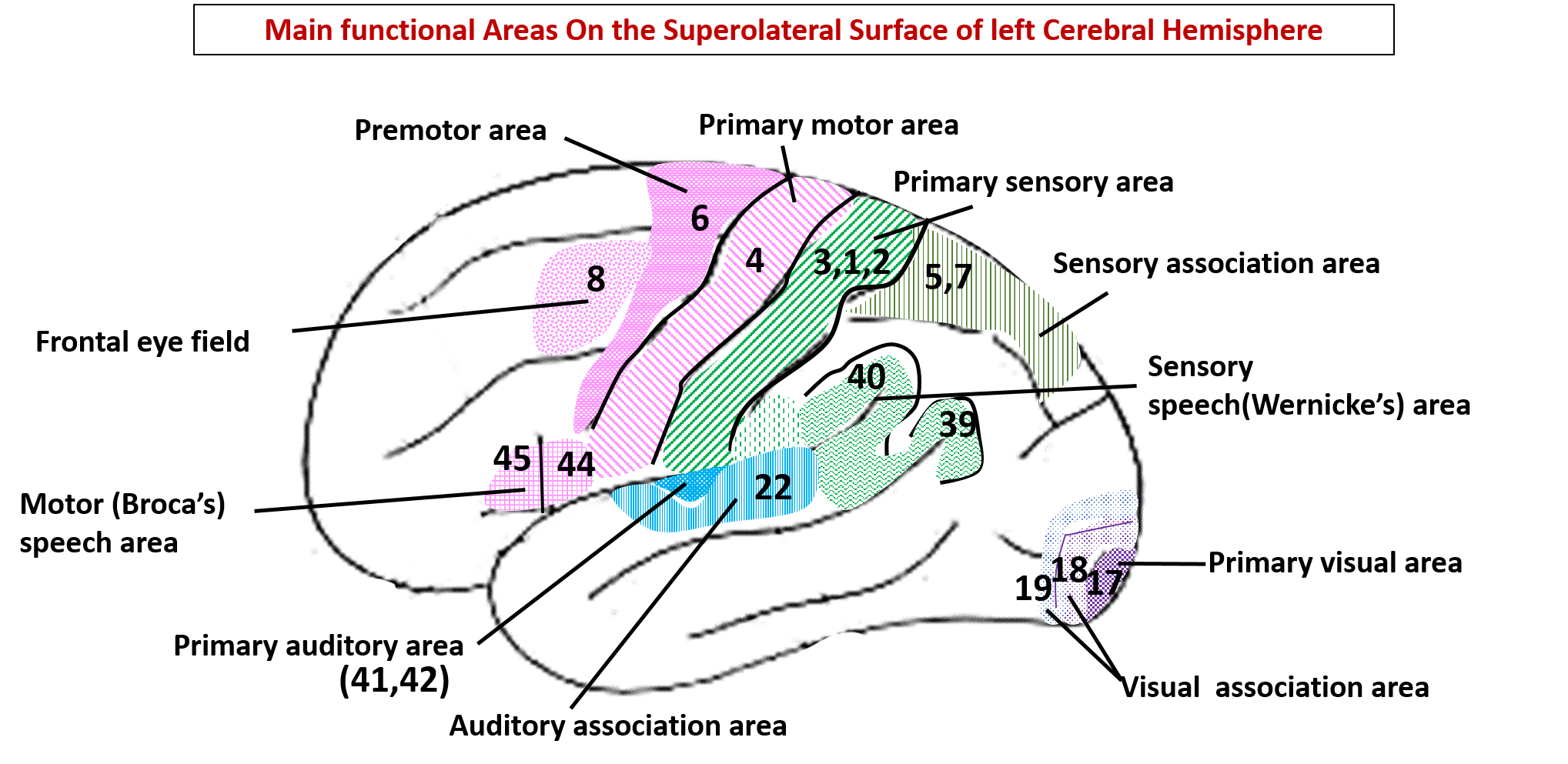
Name the functional areas located in the different lobes of cerebral hemisphere along with their function, connections and effect of lesion.
Lobe of Cerebrum Functional area Brodmann area/Gyri-sulci Connections Function Effect of lesion of the area Frontal Primary motor area (MsI) Area no. 4 ( precentral gyrus, anterior wall of central sulcus , anterior part of paracentral lobule). Afferents: premotor area, supplementary motor area, motor speech area, frontal eyefield and ventral anterior and ventral lateral nucleus of thalamus ( fibers from basal nuclie and cerebellum relay in thease thalamic nuclei). Efferents: will follow corticospinal, corticonuclear, corticoreticular, corticorubral pathways. Intiates and controls contraction of skeletal muscles of mainly contralateral side. Axila muscles and muscles of upper part of face, larynx, pharynx have bilateral control. Lesion of primaty motor cortex of one side - spastic paralysis of muscle of extremities (hemiplegia), mucles of upper part of face, larynx and pharynx are spared ( bilateral control) Frontal Premotor area Area no 6, posterior most part of superior,middle and inferior frontal gyrus. Afferents: sensory areas of cerebral cortex, ventral anterior and ventral lateral nucleus of thalamus. Efferent: To primary motor area Plan/programmeslilled motor activity on the basis of past experience and direct the primary motor area to execute the movement. Also contols extrapyramidal system. Initially spastic paralysis on contralateral side, later difficulty in performance of skilled movements. Frontal Frontal eye field Area no. 8, part of middle frontal gyrus just in front of premotor area. Afferents: from visual association area. Efferent: to contraletral nuclei of IIIrd, Ivth and Ivth cranial nerves (extraocular muscles). Causes conjugate deviation of eyes to the opposite side. Eyes deviate to the site of lesion and inability to turn the eyes to the opposite side. Frontal Prefrontal cortex Area no. 9,10,11,12. All of frontal lobe anterior to premotor area. Afferent and efferent to association cortex of general sensory area (5,7), vvisual association area (18,19) and auditory association area(22). Also coonected to dorsomedial nucleus of thalamus, hypothalamus, limbic system and cerebellum. It is concerned with individual personality. Involved in determining judgement, initiative, concentration, depth of emotion,social and moral awareness. Bilateral damage can lead to changes in behaviour- loss of judgement, initiaive and concentration, does not conform to social norms regarding behaviour, dress etc. Parietal Primary sensory area (SmI) Area no. 3,1,2. Posterior wall of central sulcus, postcentral sulcus, posterior part of paracentral lobule. Afferents: from ventral posterolateral and ventral posteromedial nucleus of thalamus. Efferent: to sensory association area, primary motor rea, prefrontal cortex and some fibers along pyramidal tract. Perception of exteroceptive (pain, temperature ,touch)and proprioceptive (vibrations, joint position and state of muscle ontraction) Loss of exeroceptive and proprioceptive sensations from the contralateral half of the body. Parietal Sensory association area Areano. 5 and 7, superior parietal lobule and precuneus. Afferents: from primary sensory area and thalamus analysis and integration of general sensationfor perception of shape size and texture of objects. It enables individuals to identify the objects placed in their hand with closed eyes. This ability is known as stereognosis. Inability to identify objects by only touching/feel - called tatile agnosia or astereognosis. Parietal Sensory sppech area of Wernike is located in dominant cerebral hemisphere (mostly the left cerebral hemisphere) Area no. 39,40. part of 22, supramarginal,angular gyri andposterior part of superior temporal gyrus. Afferents: from auditory and visual association areas. Efferent: to motor sppech area via arcuate fasciculus. Interpretation of spoken and written words and comprehension of language. Person is unable to understand both spoken and written words even though his vision and hearing are normal Temporal Primary auditory area Area no. 41,42, transverse temporal gyri (Heschl's gyri) on the superior surface of superior temporal gyrus Afferents: from medial geniculate body through aauditory radiation. Efferent: to association auditory area. To perceive the quality, pitch and loudness of voice and direction of source of sound. Lesion on one side leads to slight loss of hearning on both the sides (more on opposite side), as auditory area receives information from cochlea of both sides. Temporal Secondary (association) auditory area Area no. 22, lateral surface of superior temporal gyrus. Afferents: from primary auditory area. Efferent: to sensory speech area. Interprets sounds using past experience. Failure to interpretmeaning of words- known as word deafness or verbal agnosia Occipital Primary visual area Area no. 17, floor and walls of calcarine culcus and on superiolateral surface around the occipital pole. Macular area( area of maximum acuity)occupis posterior 1/3rd of the visual area. Afferents: From lateral geniculate body via optic radiation. Efferent: to visula association/secondary auditory area. Perceives visual impressions like colour, size, transparency, brightness etc. Loss of vision in the opposite visual field (homonymous hemianopia).Macular vision is spared as the cause of lesion is damage of posterior cerebral artery and the macular area receives blood supply from both the middle as well as posterior cerebral arteries. Occipital Secondary/association visual area Area no.18,19, surrounds the primary visual area Afferent: from primary visual area. Efferent:to sensory speech area. recognition of objects using past experience. Loss of ability to recognize objects in the oppsite visual field - known as visual agnosia

It’s really a great and beautiful platform with simplified work ..thanks ❤️